


위내시경시 간낭종에 의한 외부압박을 너무 단정적으로 GIST로 설명 들은 경우 - 동대문구 답십리, 장안동, 우리안애 우리안愛 내과
- Byoung-Yeon Jun

- 2024년 4월 23일
- 1분 분량
상기 환자 다음날 방문 (신우신염에 대해서는 다시 정리 필요, 양측!)
왼측 늑골각 압통은 많이 호전되었는데 우측 늑골각 압통이 있고 앞쪽에서도 우상복부 압통이 있어 초음파 시행
담낭이나 신장에 뚜렷한 소견은 없었으며 (신우신염의 초음파적 소견은 없다.)
Ultrasound is insensitive to the changes of acute pyelonephritis, with most patients having 'normal' scans. Abnormalities are identified in only ~25% of cases. Possible features include:
particulate matter/debris in the collecting system
reduced areas of cortical vascularity by using power Doppler
gas bubbles (emphysematous pyelonephritis)
abnormal echogenicity of the renal parenchyma
focal/segmental hypoechoic regions (in edema) or hyperechoic regions (in hemorrhage)
mass-like change
Ultrasound is, however, useful in assessing for local complications such as hydronephrosis, renal abscess formation, renal infarction, perinephric collections, and thus may guide management.
초음파 중에 과거에 **대학교 병원에서 내시경 후 CT등 시행전에 GIST 위장관기질종양이라고 단정지어 설명들었다고 확인. CT등 찍어보니 간낭종이었다.
아래와 같이 확인됨, S2 segment에 3cm 내외가 되며 위 (stomach) 의 저부 (fundus) 에 닿아 있으면 관찰될 수 있겠다.
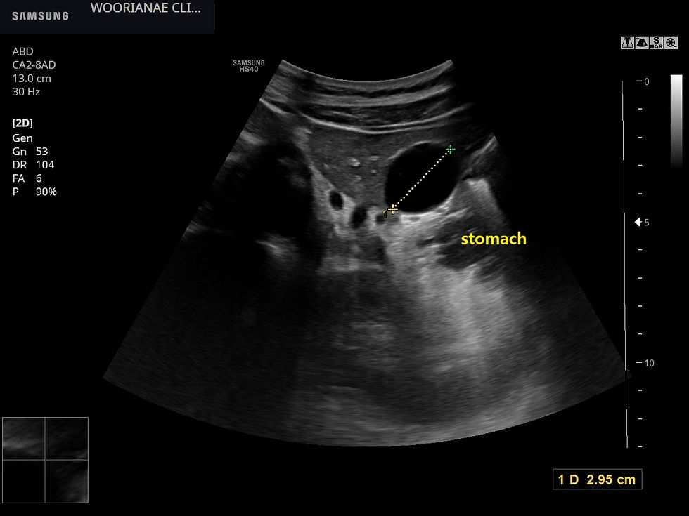
세워서 본 모습

위내시경시 점막하 종양이라고 생각되면 외부장기의 압박도 도려해야 한다.
위 하체부, 대만에서 관찰되는 3cm의 점막하 종양
....
대학병원을 갔다왔으나...
동대문구 답십리 우리안애, 우리안愛 내과, 건강검진 클리닉 내과 전문의 전병연
댓글